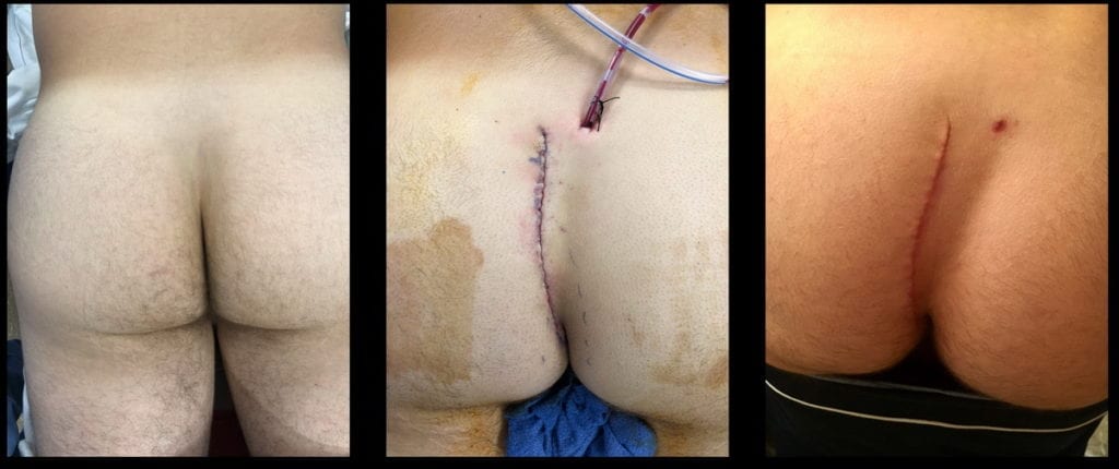
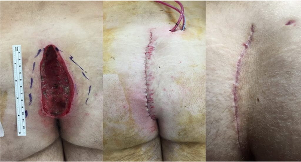
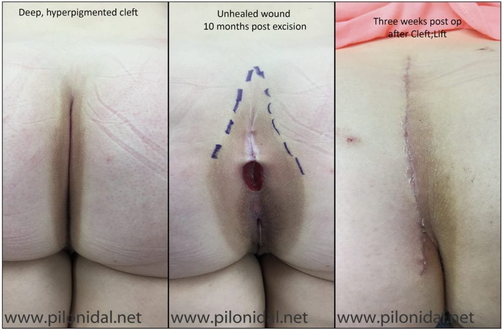
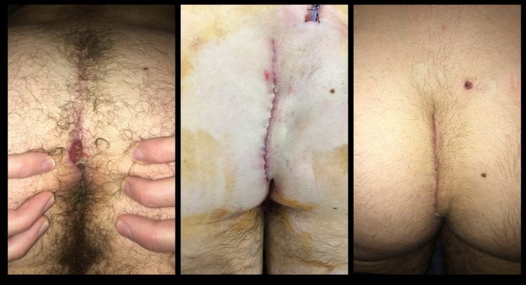
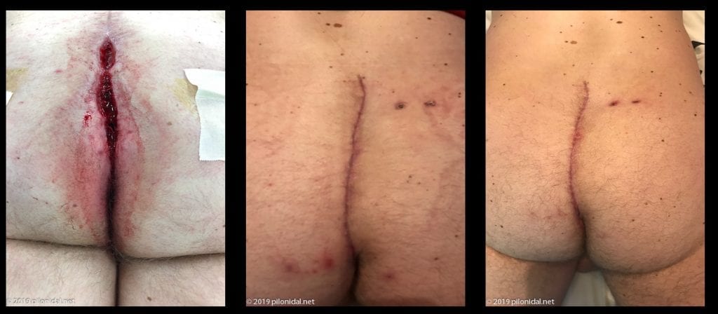
This is an example of a cleft-lift in a patient who had pain and midline pores draining pus. He had not had previous surgery. As you can see from the “after” photo, aside from the scar (which will fade) the contour is quite different, but not necessarily abnormal in appearance. This patient had 3 previous operations, the last of which left him with a 15 cm open wound. He contacted us right after the last operation and we had him come immediately to Wisconsin for a cleft-lift which has healed quickly and durably. Clicking on this image will take you to a discussion about seeing us for a cleft-lift to salvage the situation after an open excision.This patient presented with a sinus opening (seen as the spot above the cleft), and multiple midline wounds extending almost to the anus. A cleft-lift healed quickly, and the last photo is at 6 weeks post-op.This patient came to us after having a wide excision which was treated with packing and dressing changes for 10 months, but never healed completely. She subsequently came to us for a successful cleft-lift.This patient had a wide excision that failed, and then a cleft-lift (by another surgeon) which left him with an open area at the bottom, near the anus. A revision of the cleft-lift has healed without problems.This patient had had three previous wide excisions that left him with a long, open wound seen on left. The middle image is 5 weeks post-op, and the image on the right is two years post op, with no evidence of recurrent disease.14 year old male after two failed excisions. Image on the right is 9 weeks after cleft-lift surgery.This is a patient who had previously had a wide excision which did not heal. Photo on the right is 2 months post op.